

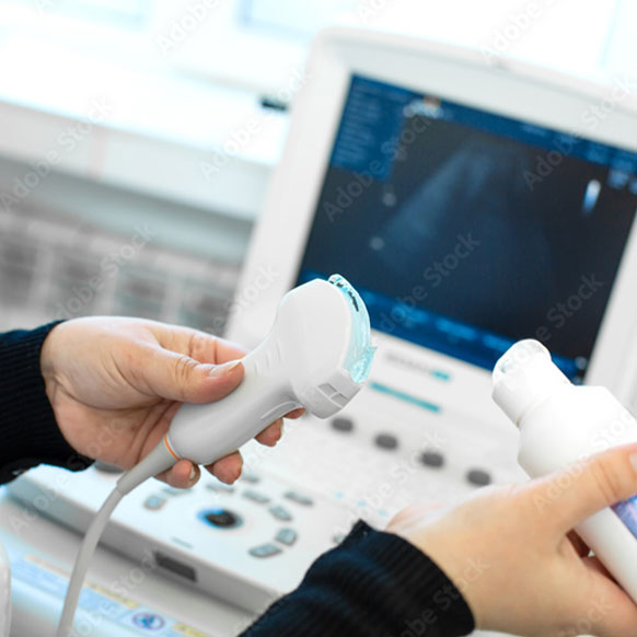
An ultrasound is a diagnostic procedure that produces images of the inside of the body on a screen (sonogram) by passing high frequency sound waves into the body. The pelvic ultrasounds is the primary radiographic method for evaluation of the female pelvis and is usually recommended to diagnose causes of pelvic pain, abnormal vaginal bleeding, infertility and other issues. This method of imagery does not subject the patient to any radiation, unlike an X-ray or CT Scan. The quality and the ability to see the pelvic organs can be more difficult in patients whom are obese.

Like the transabdominal ultrasound, the transvaginal ultrasound utilizes high frequency sound waves to project images of the inside of the body onto a computer screen. In situations were we are unable to get a good look at the pelvic organs on transabdominal scan, a transvaginal may be necessary. This method involves placing a special ultrasound probe in the vagina. The benefit of this is that it places the ultrasound probe up against the cervix and much closer to the uterus and other pelvic organs without worrying about obstruction from the abdominal wall or any fat that my be in the way. This is the primary method for looking at and dating a first trimester pregnancy since the fetus is often seen at a much earlier gestational age than with the transabdominal approach as well as evaluating the ovaries for ectopic pregnancies.
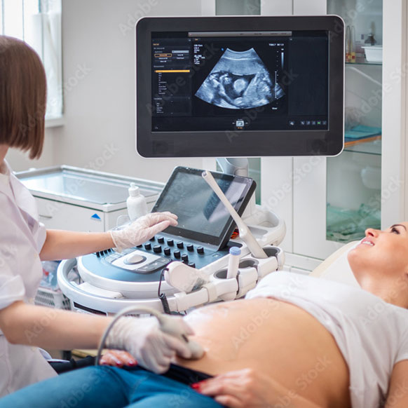
An ultrasound is the safest way to evaluate the unborn fetus at every stage of pregnancy. An ultrasound can identify a pregnancy in the uterus as soon as the 5th week of pregnancy. A heart beat can be reliably seen as soon as 6 weeks. Fetal limbs and organs are not usually seen until much later. And the determination of sex is not always accurate. The use of ultrasound in obstetrics is mainly to screen for any structural abnormalities and to assess how well the baby is growing.

We often use the ultrasound to aid in the assessment of fetal well being by doing a biophysical profile during the third trimester in those women who may have a high risk pregnancy that may lead to decrease oxygen or nutrients to the baby during pregnancy. This type of ultrasound assesses that babies movements, tone, breathing movements and the amount of amniotic fluid around the baby. In some situations the patient may also be placed on a fetal monitor to assess the pattern the fetal heart rate is making. This is called a fetal Nonstress test.,